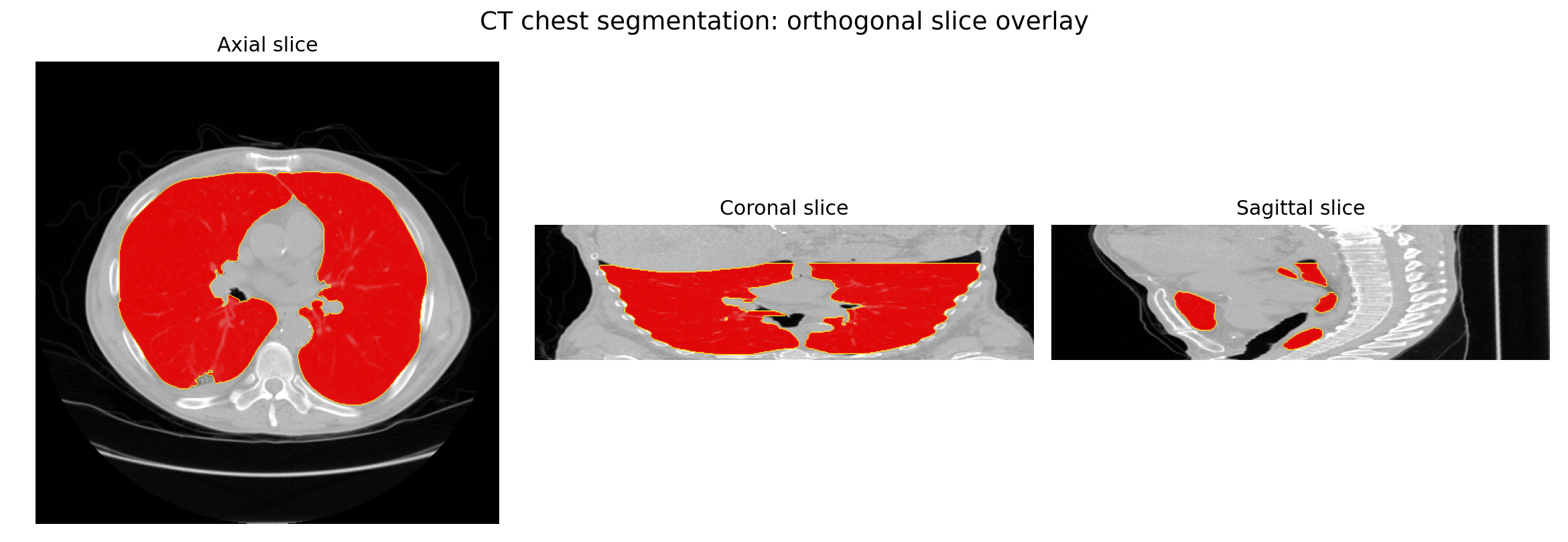
CT lung segmentation overlay across orthogonal medical image slices

Medical imaging / 3D reconstruction

3D Slicer-Compatible Segmentation Portfolio

Biomedical image-analysis samples covering MRI segmentation, CT thresholding, 3D surface reconstruction, labelmap and mesh export, measurement tables, and adjacent C. elegans neuroanatomy mapping.

Reviewer path: Start with the brain MRI and CT chest projects, inspect the screenshots and measurement tables, then use the C. elegans project as adjacent image-stack and biological structure mapping evidence.
2

3D Slicer-compatible sample segmentations

6

orthogonal and surface screenshots

4

NRRD labelmap and STL mesh exports

1

C. elegans neuroanatomy work sample

Scope note

These samples use public 3D Slicer teaching data and are portfolio demonstrations, not clinical annotations. The C. elegans project supports adjacent biomedical image-stack navigation, structure mapping, and neurobiology interpretation experience.

Featured projects

Medical image stacks to segmentations, surfaces, and measurements.

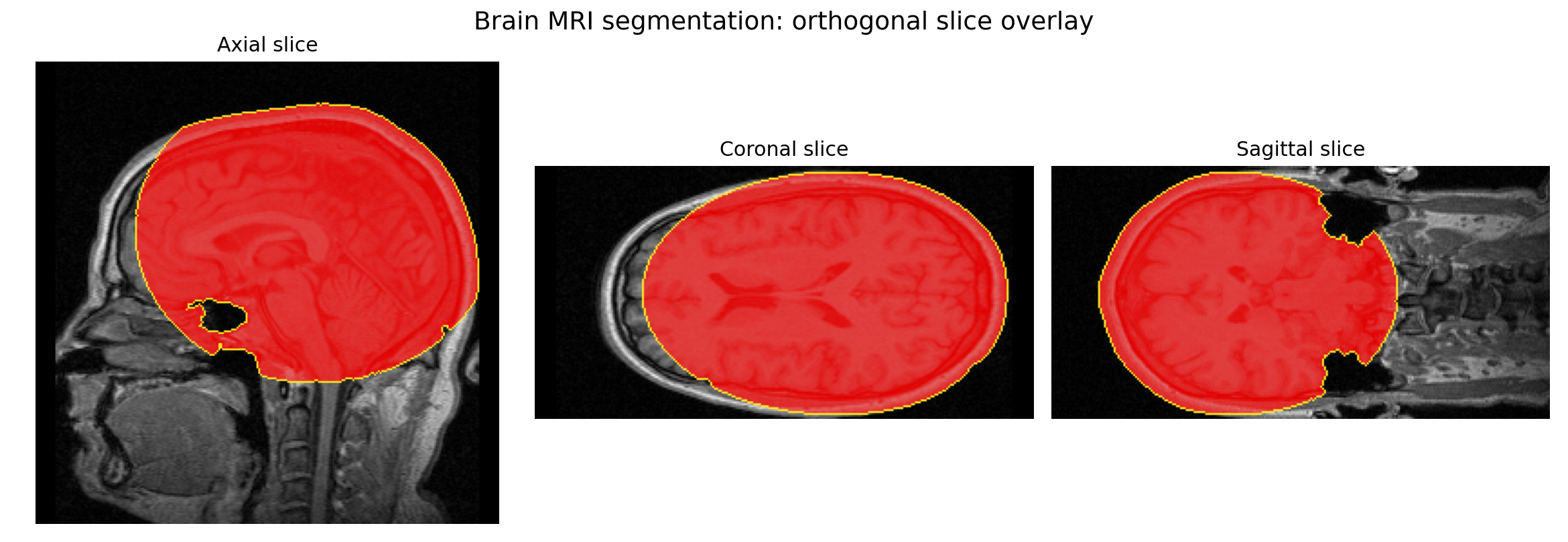
Brain MRI segmentation overlay in axial, coronal, and sagittal views
Orthogonal MRI slice overlay showing assisted brain soft-tissue ROI segmentation.

Project 1 / MRI

Brain MRI Segmentation

Assisted soft-tissue ROI segmentation from public 3D Slicer MR-head sample data. The workflow demonstrates image-stack review, ROI isolation, morphology cleanup, closed-surface generation, and measurement export.

Volume2419.338 mL
Voxels1,861,036
ExportsNRRD / STL
CT lung segmentation overlay in axial, coronal, and sagittal views
CT chest threshold segmentation isolating lung airspace from the surrounding body volume.

Project 2 / CT

CT Chest Lung Segmentation

HU threshold-based segmentation from public 3D Slicer CT-chest sample data. The workflow demonstrates lung airspace isolation, border/background air removal, connected-region cleanup, 3D surface reconstruction, and volume measurement.

Volume6129.845 mL
Voxels4,225,907
ExportsNRRD / STL
Figure 3 presentation cover for C. elegans odr-10 and AWA neuron-related analysis
Figure 3 work sample from C. elegans odr-10/AWA neuron-related behavior analysis.

Project 3 / Adjacent biomedical reconstruction

C. elegans Neuroanatomy Mapping with IMOD/3dmod

Adjacent biomedical image-analysis experience from a Harvard Medical School-affiliated C. elegans neurobiology context. The work involved biological structure mapping, microscopy/image-stack interpretation, and C. elegans odr-10/AWA neuron-related behavior analysis.

Image-stack navigation Manual/assisted tracing Neuron/glia morphology Biological structure mapping

Workflow capability

The workflow mirrors a practical 3D Slicer asset pipeline.

Load

Open volumetric NRRD sample data and inspect axial, coronal, and sagittal planes.

Segment

Use thresholding, ROI constraints, connected components, hole filling, and morphology cleanup.

Reconstruct

Generate closed-surface models from labelmaps with marching-cubes style surface extraction.

Export

Prepare reviewer-ready labelmaps, STL meshes, screenshots, and measurement tables.

Document

Write project notes that distinguish public teaching data, portfolio scope, and biological context.

Evidence package

Project files linked for technical review.

The two 3D Slicer-compatible segmentation samples live in standalone GitHub repositories. C. elegans is included as adjacent biomedical image-analysis context, not as a separate repository.

Asset Format Purpose
Brain MRI standalone reporepoSegmentation notes, screenshots, NRRD labelmap, STL mesh, and measurements
CT chest standalone reporepoThresholding workflow notes, screenshots, lung labelmap, STL mesh, and volume table
Heiman Lab contextcontextAdjacent C. elegans nerve cell, glia, microscopy, and morphology research background
C. elegans Figure 3 analysis contextcontextodr-10/AWA neuron-related food-search behavior analysis used as adjacent biomedical image-analysis context

Survey wording: 3D Slicer-compatible portfolio samples for brain MRI segmentation, CT chest/lung threshold segmentation, 3D surface reconstruction, STL/NRRD export, and volume measurement tables, plus adjacent C. elegans neuroanatomy/image-stack mapping experience with IMOD/3dmod-style workflows.